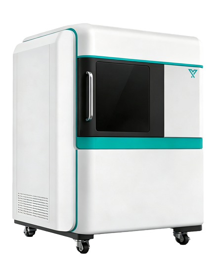
1. High‑sensitivity scientific‑grade cooled camera
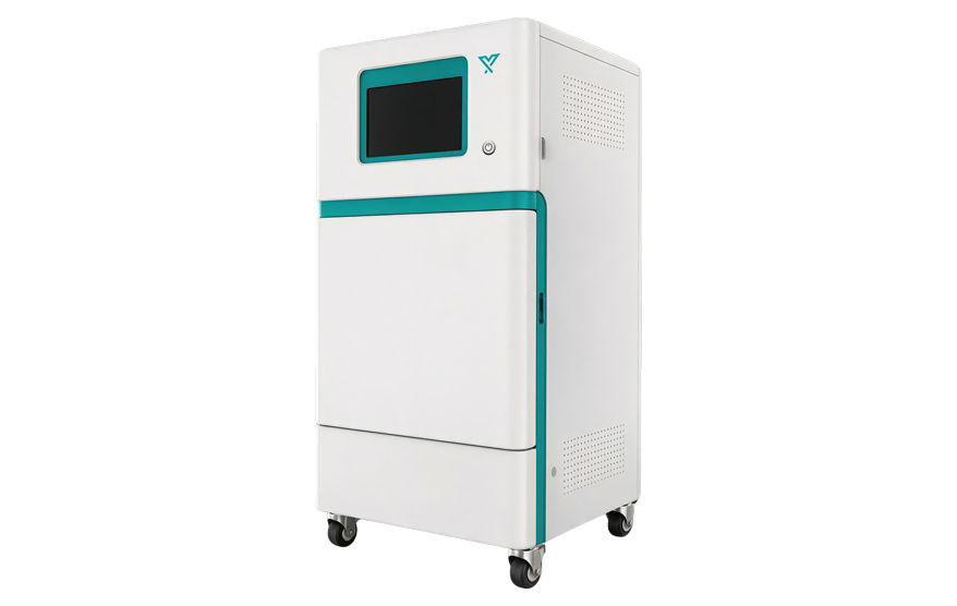
This system adopts a 1‑megapixel, 16‑bit scientific‑grade cooled camera, balancing faint‑signal detection and spatial detail representation. Deep cooling effectively reduces dark current and long‑exposure noise, making it suitable for detecting low‑expression reporter genes and weak bioluminescent samples.
2. Bioluminescence imaging module
The fully enclosed dark chamber, paired with a large‑aperture low‑loss imaging optical path, detects faint photon signals generated by Luciferase reporter genes and other bioluminescent reactions. It supports exposures ranging from seconds to minutes, and allows adjustment of pixel binning, aperture and exposure time according to sample signal intensity.
3. Multi‑band fluorescence excitation module
Equipped with multi‑channel LED excitation light sources covering ultraviolet, blue, green, red and near‑infrared bands. It is compatible with GFP, YFP, RFP, chlorophyll, fluorescent dyes and other plant fluorescent markers.
Each excitation channel can be independently controlled by software to meet experimental requirements for single‑channel, multi‑channel and combined detection of different fluorescent markers.
4. Motorized emission filter system
The 24‑position motorized filter wheel automatically switches emission channels according to imaging protocols. Narrow‑band filters minimize excitation‑light leakage and crosstalk between different fluorescent signals, improving the identification capability of target signals.
5. Chlorophyll autofluorescence subtraction
By utilizing spectral differences across different wavelength bands, the software separates target fluorescence from chlorophyll autofluorescence. It reduces interference from intrinsic plant‑tissue fluorescence on the detection of GFP, RFP and exogenous fluorescent probes.
6. Motorized lifting sample stage
The motorized lifting sample stage enables automatic switching among different fields of view. Users can flexibly switch between high‑resolution local imaging of seeds and leaves, and large‑field‑of‑view imaging of petri dishes, microplates and whole plants.
7. Compatibility with multiple types of plant samples
The imaging chamber accommodates petri dishes, microplates, potted seedlings, detached leaves and other plant samples. Optional accessories include sample holders of varying heights, fixing plates for culture vessels and root‑imaging attachments.
8. Time‑lapse acquisition and quantitative analysis
The software supports auto‑exposure, auto‑focus, continuous acquisition and time‑series experiments. It enables ROI drawing, and analysis of total signal intensity, mean intensity, signal area, signal‑to‑background ratio, as well as variation trends across different time points.