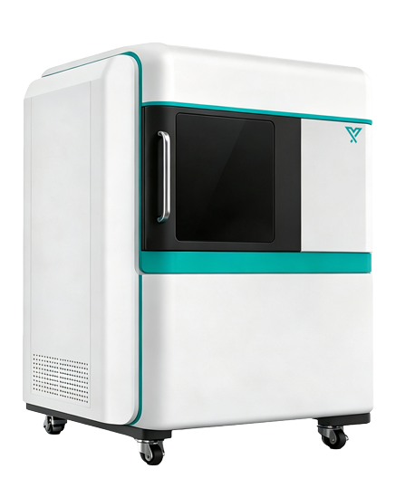
1. High‑sensitivity cooled camera
Adopts a ~1‑megapixel, 16‑bit scientific‑grade cooled camera. Its large‑pixel design delivers strong photon‑collection capability. Combined with deep‑cooling technology to reduce dark current and long‑exposure noise, it is suitable for detecting faint bioluminescent signals in deep tissues or low‑expression models.
2. High‑throughput imaging optical path
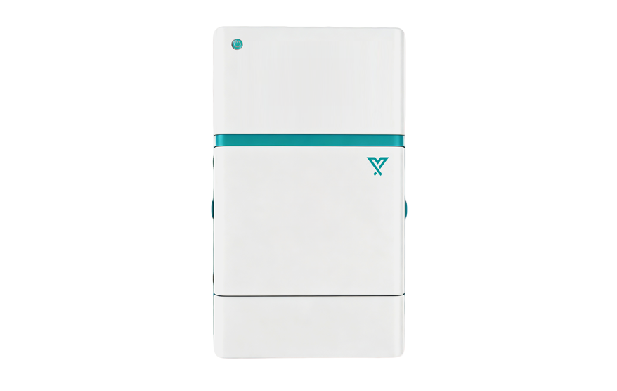
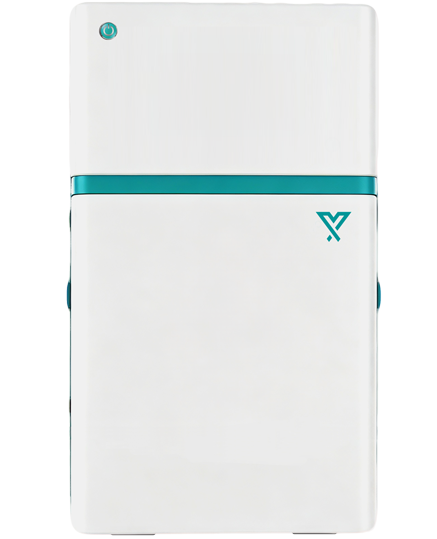
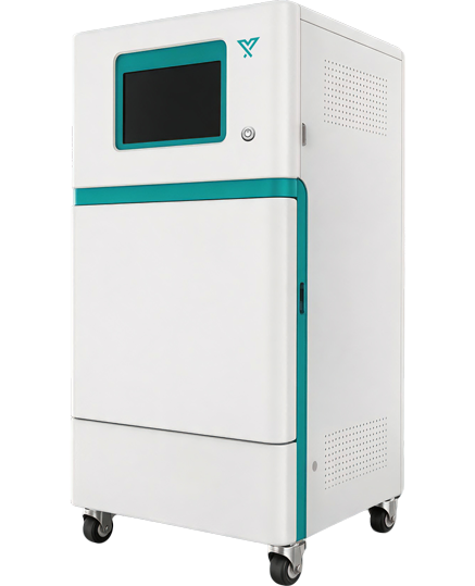
Equipped with a large‑aperture motorized lens and low‑loss optical components to boost the transmission and collection efficiency of limited photons. The fully enclosed dark chamber minimizes ambient‑light interference, providing a stable dark environment for exposures ranging from seconds to minutes.
3. Motorized stage and multi‑field‑of‑view options
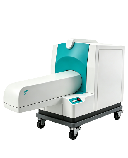
The motorized lifting stage enables automatic switching among different imaging fields of view, allowing flexible selection between high‑resolution regional imaging and whole‑body imaging of multiple animals. The maximum field of view supports simultaneous imaging of up to five mice, improving the efficiency of batch experiments.
4. Animal temperature maintenance and anesthesia support
The stage features temperature‑control function to sustain animal body temperature during imaging. The system can be connected to an inhalation gas anesthesia device, with multi‑channel nose cones delivering anesthetic gas to multiple animals concurrently.
5. Anesthetic waste‑gas recovery
Fitted with active or passive waste‑gas recovery ports to collect residual anesthetic gas inside the imaging chamber. This reduces exposure for laboratory personnel and maintains stable anesthetic depth for animals.
6. Intelligent acquisition and quantitative analysis
The software supports auto‑exposure, auto‑focus, continuous exposure and time‑series acquisition. It enables ROI drawing, calculation of total photon flux, average radiance intensity, signal area and inter‑group difference analysis, as well as longitudinal comparison of data across different time points.